Canonical Question
Procedure – Venous Access
Master answer
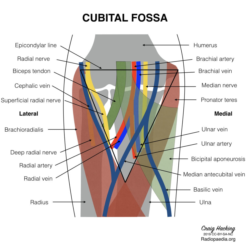
Antecubital fossa – a triangular view on the anterior aspect of the elbow
Boundaries
- Medial – lateral border of pronator teres from the medial epicondyle
- Lateral – medial border of brachioradialis from the lateral supraepicondylar ridge of the humerus
- Superior – an imaginary horizontal line connecting the medial and lateral humeral epicondyles
- Inferior – the apex is directed inferiorly and is formed by the meeting point of the medial and lateral boundaries
- Deep (floor) – brachialis (proximal) and supinator (distal) muscles
- Superficial (roof) – skin, superficial fascia (containing the medial cubital vein, lateral and medial cutaneous nerves of
- the forearm, deep fascia (reinforced by the bicipital aponeurosis)
Contents (from medial to lateral)
- Median nerve – leaves the fossa between the two heads of pronator teres
- Brachial artery – bifurcates into the radial and ulnar arteries at the apex of the fossa
- Biceps tendon
- Radial nerve – not strictly in the fossa but is in the vicinity, passing underneath brachioradialis
Venous anatomy in the superficial fascia of the fossa
(from medial to lateral)
- Basilic vein
- commences in the medial forearm, joined by the medial cubital vein at the level of the ACF and then perforates the brachial fascia above the medial epicondyle, joining the brachial veins to form the axillary vein.
- Favoured for PICC cannulation because it is usually of substantial size, with predictable anatomy and is easily viewed with USS.
- Medial cubital vein (variable anatomy)
- Cephalic vein
- commences in the lateral forearm and continues up the lateral aspect of the arm before it enters the deltopectoral groove and empties into the axillary vein.
- Less used for PICC insertion because the presence of the claviopectoral fascia superiorly causes it to enter the axillary vein at an acute angle, which is often difficult for the catheter to negotiate.
Gladwin 2016
Exam appearances
| Exam | Exact wording | Relationship | Success |
|---|---|---|---|
| 2012B Q07 | Describe the anatomy of the antecubital fossa and peripheral veins of the upper arm relevant to a peripherally inserted central venous catheter (PICC). | historical_member | — |