Canonical Question
Procedure – Arterial Access
Master answer
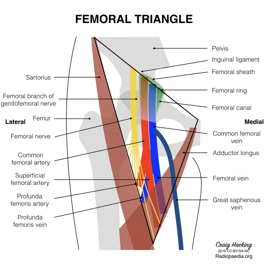
Femoral triangle (aka Scarpa’s triangle)
- The major boundaries can be recalled with the mnemonic SAIL:
- lateral border: medial border of sartorius
- medial border: medial border of adductor longus
- superior border: inguinal ligament
- floor: iliopsoas (laterally) and pectineus (medially)
- roof: skin, subcutaneous tissue, a continuation of Scarpa’s fascia, great saphenous vein (joins the femoral vein), superficial lymph nodes, fascia lata
- Contents (From lateral to medial):
- femoral nerve
- femoral sheath (thickening of the deep fascia of the thigh) which has three compartments (from lateral to medial):
- femoral artery and its branches (within the lateral compartment of the femoral sheath) and the femoral branch of the genitofemoral nerve
- femoral vein (within the intermediate compartment of the femoral sheath) and deep lymph nodes
- femoral canal (the medial compartment of the femoral sheath) which contains fat and lymph nodes (of Cloquet)
Femoral sheath
- Situated within the triangle.
- Formed from the fusion and invagination of the psoas and transversalis fascia.
- Contains femoral artery, vein and tributaries, and femoral canal
Femoral artery
Main artery of the lower limb, originates as a continuation of the external iliac artery at the level of the inguinal ligament.
Course:
- Enters the femoral triangle deep to the midpoint of the inguinal ligament (clinically the femoral pulse is usually found midway between the ASIS and pubic tubercle), lateral to the femoral vein
- Passes through the triangle and exits at its apex, entering the adductor canal (Hunter’s canal)
- Exits the adductor canal by passing through the adductor hiatus in adductor magnus (at the level of the junction between the middle and lower third of the thigh) to become the popliteal artery.
- Several branches – largest is profunda femoris, the chief artery to the thigh, which arises from the lateral aspect of femoral artery, 2-5cm below the inguinal ligament. Other branches include the superficial epigastric, superficial iliac circumflex and superficial and deep external pudendal arteries.
Relationships:
- Medial – femoral vein (upper part of the triangle)
- Lateral – femoral nerve, lumboinguinal nerve
- Superior – inguinal ligament
- Posterior – posterior part of femoral sheath, pectineal fascia, psoas major tendon, pectineus and adductor longus
- Anterior – skin, superficial fascia lata, lymph nodes, anterior part of femoral sheath
Sources: Gray’s Anatomy, Radiopedia
JC 2019
Exam appearances
| Exam | Exact wording | Relationship | Success |
|---|---|---|---|
| 2010A Q23 | Describe and or illustrate the anatomy relevant to the insertion of an arterial line into the femoral artery | historical_member | — |