Canonical Question
CVS – Excitation
Master answer
Sodium channels
Classified into:
- Voltage-gated sodium channels
- In exitable cells such as myocytes, neurons, certain glia
- Very selective to Na+
- Responsible for rising phase of action potentials
- Triggered by change in membrane potential (voltage)
- Ligand-gated sodium channels
- NMJ – Nicotinic receptors (ACH – ligand)
- Permeable to Na+ and K+
- Triggered by binding of ligands to channel
- Leak sodium channel
- Ungated, always open
- Contributes to depolarization from K+ potential
- Increasing permeability lowers RMP, brining it closer to trigger of an action potential
Voltage-gated Sodium Channel
- Family consisting of Nav 1.1-1.9 encoded by genes SCN1A to SCN9A respectively
- Human Cardiac Sodium channel Nav 1.5 encoded by SCN5A
- responsible for the generation of the rapid upstroke of the myocardial action potential
- determines impulse conduction velocity in cardiac tissue (affects action potential upstroke velocity — extent of intercellular communication via gap junctions)
- Mutations in the gene encoding this channel (SCN5A) linked to three forms of primary electrical disease (channelopathies)
- the long QT syndrome (LQTS)
- the Brugada syndrome (BS) and
- cardiac conduction defects such as broad QRS, LBBB/RBBB, prolonged PR intervals, and other channelopathies like familial AF and idiopathic VF.
Structure
composed of an α subunit (forms ion conduction pore) and one or more auxiliary β subunits (several functions incl. modulation of channel gating)
α subunit
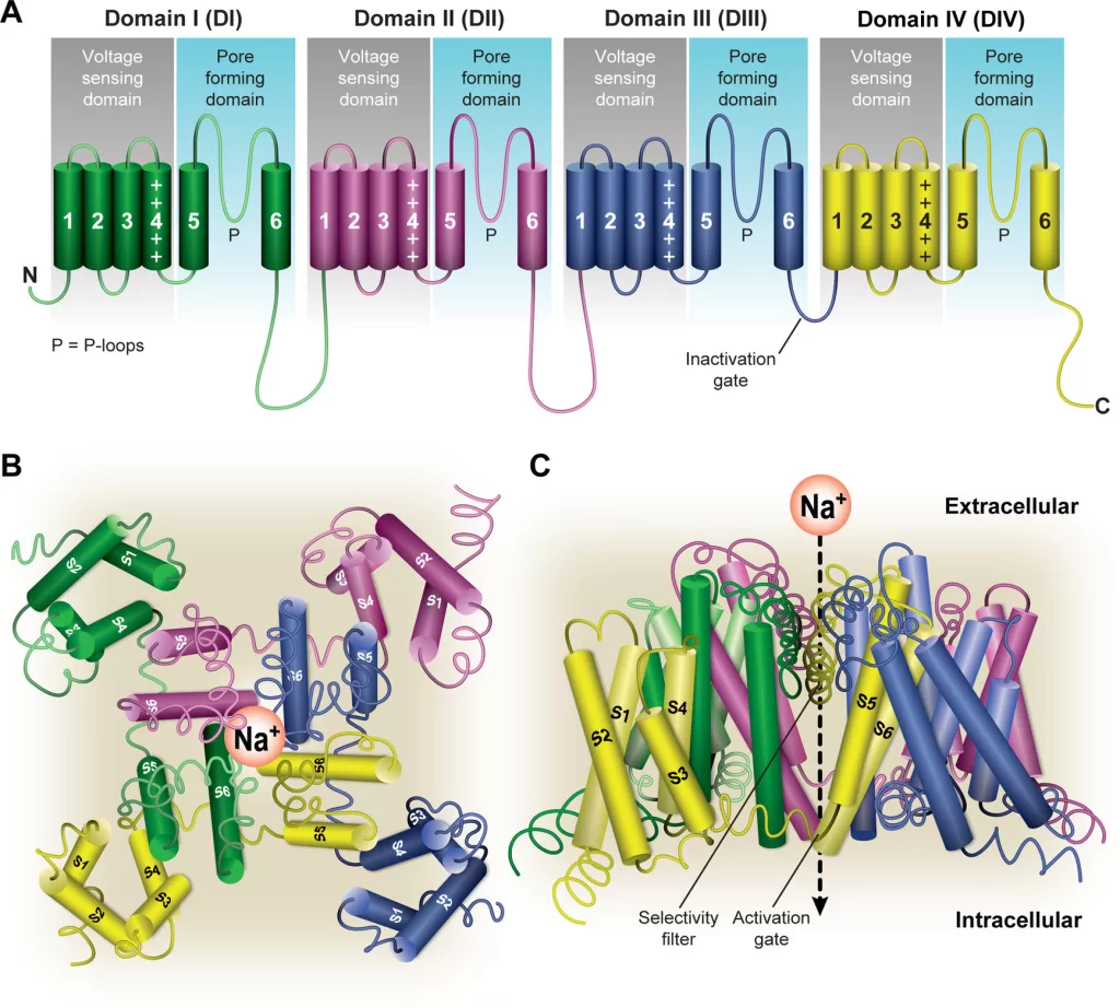
- α subunit is the largest subunit and contains both the pore region of the channel and the voltage sensor
- forms core of the channel and is functional on its own
- indepently able to form a pore in the cell membrane that conducts Na+ in a voltage-dependent way
- When accessory proteins assemble with α subunits, the resulting complex can display altered voltage dependence and cellular localization.
- Principal α-subunit composed of four homologous domains (DI–DIV), each containing six transmembrane segments (S1–S6). The four domains are attached to one another by cytoplasmic linker sequences.
- Highly conserved S4 segment acts as the channel’s voltage sensor
- When stimulated by a change in transmembrane voltage, this segment moves toward the extracellular side of the cell membrane, forming the central pore cavity and allowing the channel to become permeable to ions
- Central Pore Cavity
- Conduct ions
- External portion(i.e., more extracellular): formed by the “P-loops” (the region between S5 and S6) of the four domains. This region is the most narrow part of the pore and is responsible for its ion selectivity (Selectivity filter). Keep out anions and large cations (K+)
- Inner portion (i.e., more cytoplasmic): is the pore gate and is formed by the combined S5 and S6 segments of the four domains.
- Also features lateral tunnels or fenestrations: that run perpendicular to the pore axis. Connect the central cavity to the membrane. Proposed to be important for drug accessibility.
Gating
- have two gates
- Activating gate (m) (voltage-dependent): Opening of the activating gate allows the influx of sodium and cell depolarization.
- Inactivation gate (h) (time-dependent): closing of the inactivation gate will stop the flow of sodium regardless of the status of the activation gate
- Highly conserved S4 segment acts as the channel’s voltage sensor
- When stimulated by a change in transmembrane voltage, this segment moves toward the extracellular side of the cell membrane, forming the central pore cavity and allowing the channel to become permeable to ions
- The cytoplasmic linker between domains III and IV of the α subunit is responsible for inactivation of the Na channel.
- After the channel opens, this segment of the protein is drawn to the inner surface of the channel like a “hinged lid,” – physically wedges the pore gate shut – blocks Na+ ions from passing through the pore
- 3 main conformational states: open, closed, inactivated
- Ion permeable only in open state
- Open: m gates open rapidly
- Closed(resting): m closed, h open
- Inactive:
- Transitions:
- Activation/Deactivation (Closed ⇌ Open)
- Inactivation/Reactivation (Open ⇌ Inactivated)
- Recovery from inactivation/closed-state inactivation (Inactivated ⇌ Closed)
Ionic Events
| Action Potential | Membrane Potential | Target Potential | Gates | NA+ / RP | Gate’s Target State | Phase of Cardiac AP |
|---|---|---|---|---|---|---|
| Resting | −90 mV | −70 mV | m closed | Ready to go | Deactivated → Activated | Phase 4 |
| Rising | −70 mV | 0 mV | m open rapidly (0.1-0.2sec) | Na flow in | Activated | Phase 0 |
| Rising | 0 mV | +20 mV | h starts closing | Na flow slows | Activated → Inactivated | Phase 0 |
| Falling | +20 mV | 0 mV | h closed (takes 10msec) | Na+ channel refractory period starts | Inactivated | Phase 1 |
| Falling | 0 mV | −90 mV | h remains closed | RP ongoing | Inactivated | Phase 2,3 |
| Undershot | −90 mV | −95 mV | h open, m closes | RP ends | Inactivated → Deactivated | Phase 4 |
| Rebounding | −95 mV | −90 mV | m closed | ready to go | Deactivated | Phase 4 |
β subunit
- Four different protein isoforms
- each about one-tenth of the mass of the α subunit
- anchored in the cell membrane through one transmembrane segment
- Extracellular domains are large and have structures similar to those of immunoglobulins.
- Regulate the expression and subcellular targeting of Na channels, and modulate the function (gating) of the α subunit
Exam appearances
| Exam | Exact wording | Relationship | Success |
|---|---|---|---|
| 2022B Q05 | Outline the structure of fast cardiac sodium channels and describe in detail how they work. | historical_member | — |