Canonical Question
CVS – Excitation
Master answer
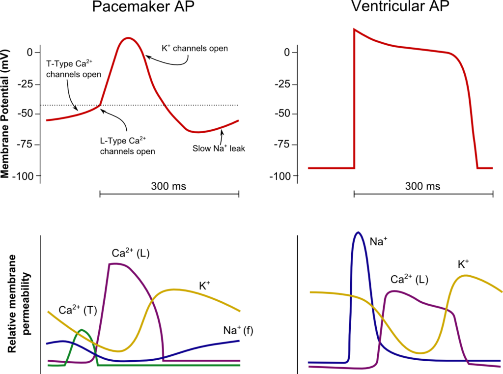
SA Node
Ventricle
Resting membrane potential
- No set RMP, however approx. -60mV
- βadrenergic stimulation causes a less negative RMP and increased slope of the initial upstroke
- muscarinic stimulation causes a more negative RMP and a decreased slope of the initial upstroke
Resting membrane potential
- Approx -90mV
Threshold potential
- Approx 40mV
Threshold potential
- Approx. -50mV
Phase 0
- Funny Na+ current allow leakage of Na+ into the the cell slowly increasing membrane potential to the threshold potential
T and L-type Ca2+ channels (voltage gated) open when threshold potential reached
- Ca2+ influx
- Depolarization
- Shallow upstroke compared to ventricular myocyte
Phase 0
- Rapid depolarization
- Opening of fast Na+ channels
- Influx of Na+
- Overshoot to +20mV
Phase 1
- Early repolarization
- Closure of fast Na+ channels and opening of K+ channels (transient outward, inward rectifier)
- Efflux of K+
Phase 2
- Plateau
- Opening of intially T-type Ca2+ channels and subsequently L-type Ca2+ channels
- Ca2+ influx balances K+ efflux
- Na+ channels in closed state à absolute refractory period
- No action potential can be generated in this state
Phase 3
K+ channels open and Ca2+ channels slowly close
- K+ efflux
- Repolarization
- No plateau phase
Phase 3
- Repolarization
- Closure of Ca2+ channels while K+ channels remain open (delayed rectifier)
- K+ efflux
- Returns potential towards RMP
- Na+ channels transition to inactive state à relative refractory period
- Another action potential can be generated with greater adequate stimulus
Phase 4
- Na+/K+ ATPase and Na+/Ca2+ ATPase maintain ionic gradients
Phase 4
- RMP
- Na+/K+ ATPase and Na+/Ca2+ ATPase maintain ionic gradients
Sakurai 2016
Exam appearances
| Exam | Exact wording | Relationship | Success |
|---|---|---|---|
| 2017B Q21 | Describe and compare the action potentials from cardiac ventricular muscle and the sinoatrial node | historical_member | — |
| 2013A Q19 | Describe and compare the action potentials from cardiac ventricular muscle and the sinoatrial node. | historical_member | — |
| 2016B Q11 | Compare the action potentials of a sino-atrial node cell and a myocardial cell. | historical_member | — |
| 2020B Q01 | Describe and compare the action potentials from cardiac ventricular muscle cells and the sino-atrial node. | historical_member | — |
| 2025B Q03 | (a) Describe the action potential of the following: (i) the sinoatrial node (35% of marks) (ii) the cardiac ventricular muscle cells (35% of marks) Include in your answer the phases and ionic events involved with each action potential. (b) Explain the implications of the differences between these action potentials (30% of marks). | safe_repeat | 83.00% |