Canonical Question
VQ – Dead space
Master answer
Dead space
- Ventilated lung volume in which no gas exchange occurs
- Anatomical dead space
- Dead space volume of conducting airways
- Oropharynx, nasopharynx and first 16 divisions of the tracheobronchial tree
- Approx 2ml/kg
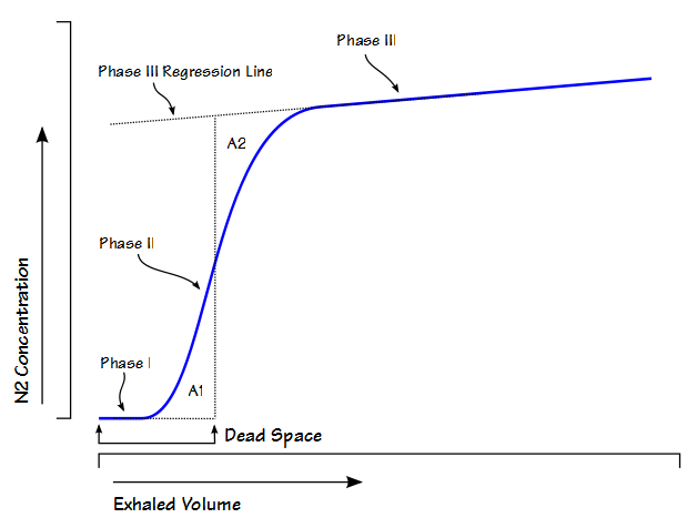
- Measured by fowlers method (N2 washout)
- Dead space volume of conducting airways
- Alveolar dead space
- Dead space volume of parts of the respiratory airways that are ventilated but not perfused
- West Zone 1 (PA > Pa)
- Low cardiac output
- Positive pressure ventilation
- Posture
- Disease states
- Pulmonary Embolism
- West Zone 1 (PA > Pa)
- Measured by subtracting anatomical deadspace from physiological dead space
- Dead space volume of parts of the respiratory airways that are ventilated but not perfused
- Physiological dead space
- Alveolar + anatomical dead space
- Calculated from Bohr equation
- Normal arterial to end-tidal CO2 gradient is approx 5mmHg
- Increases if increased dead space
- Apparatus dead space
- Dead space contributed by artificial airway devices (endotracheal tubes, LMA, HME filters)
- Usually reduces anatomical deadspace (due to bypass of oropharynx)
Calculation / Measurement of Dead Space
Bohr’s Method
- Bohr’s Equation:
\[ {{V_D} \over {V_T}} = { {V_T \; – \; V_A} \over {V_T}} \]
- Based on the principle that all CO2 exhaled must come from ventilated alveoli
\[ {{V_D} \over {V_T}} = { {PA_{CO_2} \; – \; P\bar{E}_{CO_2}} \over {PA_{CO_2}}} \]
- PĒCO2 is mixed-expired CO2 in an expired tidal breath
- Alveolar PCO2 is difficult to measure, so the Enghoff modification is used
- which assumes PACO2 = PaCO2
\[ {{V_D} \over {V_T}} = { {Pa_{CO_2} \; – \; P\bar{E}_{CO_2}} \over {Pa_{CO_2}}} \]
Fowler’s Method
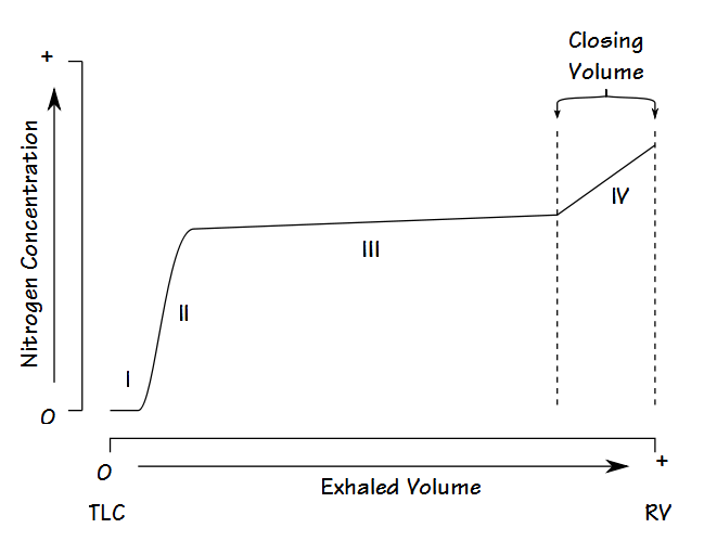
Single-breath nitrogen washout test
- Single Vital Capacity Breath – 100% O2 → Exhales to Residual volume
- Expired Nitrogen concentration and volume is measured
- Plot of concentration by volume generated
- Phase I: Pure dead space. No Nitrogen present.
- Phase II: Midpoint is volume of Anatomical dead space.
- Phase III: Expired N2 plateaus
- Phase IV: Closing capacity. Sudden Increase in Nitrogen.
Consequences of increasing dead space
- Alveolar ventilation
- RR x (tidal volume – dead space)
- Therefore, as dead space approaches tidal volume, alveolar ventilation approaches zero, and cannot be compensated by increasing respiratory rate
- PaCO2 is inversely proportional to alveolar ventilation → PaCO2 increases as alveolar ventilation decreases
- Decreased pH
- Increased central and peripheral stimulation of ventilation → Reduction in PaCO2
- Central chemoreceptors activate medullary vasomotor centre → increased sympathetic discharge
- If PaCO2 increases, alveolar CO2 in non-dead space alveoli will increase
- PAO2 will decrease according to alveolar gas equation
Sakurai / JC 2016
Exam appearances
| Exam | Exact wording | Relationship | Success |
|---|---|---|---|
| 2010A Q19 | Describe the types of dead space in the Firatory system (50% marks). Explain the consequences of increased dead space on gas exchange (50% mark) | historical_member | — |
| 2018A Q03 | Define dead space and its components (30% of marks). Explain how these may be measured (35% of marks) and describe the physiological impact of increased dead space (35% of marks). | historical_member | — |
| 2025B Q02 | (a) Define dead space and its components (15% of marks). (b) Describe the factors that affect each component (30% of marks). (c) Outline the physiological impact of increased dead space (15% of marks). (d) Describe methods of measurement of dead space (40% of marks). | safe_repeat | 34.00% |